Dr. Yang's lab reported a new BoneClear method for the robust immunolabeling of diverse cellular structures in the intact mouse bones for the first time in the field.
Conventional approaches to analyze cellular structures in the bone tissues have depended mainly on tissue sections. However, such methods obscure specific cellular structures, e.g., neural innervations, that would be better observed with the intact, unsectioned bones. In addition, histological sectioning is challenging, if not impractical, for many bone tissues such as vertebral column, skull, and limbs.
Notably, despite the recent advance of 3D fluorescence imaging methods, a whole-tissue immunolabeling procedure to detect endogenous epitopes in the bone tissues, and at the same time, to be compatible with optical clearing, has been highly desired but yet been successfully attempted.
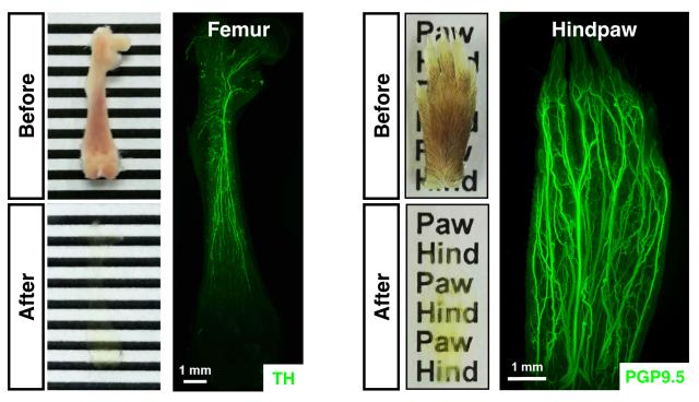
In the study online published in Cell Research (https://doi.org/10.1038/s41422-019-0217-9), Dr. Yang's lab at the IDG/McGovern Institute for Brain Research at Peking University reported a new BoneClear method for the robust immunolabeling of diverse cellular structures in the intact mouse bones for the first time in the field. With this imaging power, Dr. Yang's lab visualized the neural innervations, glial cells, blood vessels, lymphatic vessels, immune cells, and chondrocytes in the unsectioned femur, vertebral column, skull or hindpaw of adult mice. Moreover, this new method enabled the whole-tissue 3D assessment of traumatic injury- or chemotherapy-induced neuropathy in the mouse femurs and hindpaws. These results have demonstrated the unparalleled strength of the BoneClear method for the 3D fluorescence imaging of the bone tissues, which is poised to serve the research field in future investigations.